The AAPG/Datapages Combined Publications Database
AAPG Bulletin
Figure
AAPG Bulletin; Year: 2021; Issue: August DOI: 10.1306/02262118214
Return to Full Text
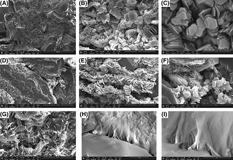
Figure 10.
Three-dimensional scanning electron microscopy (SEM) images of microporous clay minerals observed from air-dried core samples of the Cypress Sandstone. All images are secondary electron (SE) unless otherwise stated. (A) Pore-filling kaolinite. (B) Backscattered electron (BSE) image of pore-filling kaolinite vermicules. (C) Pore-filling kaolinite books. (D) Inherited chlorite-rich rims. (E) Elongated pore-filling chlorite clusters. Note face-to-edge contacts between individual blades. (F) Pore-filling chlorite clusters intergrown with blocky kaolinite. (G) Pore-filling intertwined needles of illite. (H) Pore-bridging ribbons of hairy illite. (I) Increased magnification (mag) of (H) showing individual hairs of illite latching to quartz surfaces. det = detector; ETD = Everhart-Thornley detector; HV = high voltage; LFD = large field detector; WD = working distance.
Figure 10. Three-dimensional scanning electron microscopy (SEM) images of microporous clay minerals observed from air-dried core samples of the Cypress Sandstone. All images are secondary electron (SE) unless otherwise stated. (A) Pore-filling kaolinite. (B) Backscattered electron (BSE) image of pore-filling kaolinite vermicules. (C) Pore-filling kaolinite books. (D) Inherited chlorite-rich rims. (E) Elongated pore-filling chlorite clusters. Note face-to-edge contacts between individual blades. (F) Pore-filling chlorite clusters intergrown with blocky kaolinite. (G) Pore-filling intertwined needles of illite. (H) Pore-bridging ribbons of hairy illite. (I) Increased magnification (mag) of (H) showing individual hairs of illite latching to quartz surfaces. det = detector; ETD = Everhart-Thornley detector; HV = high voltage; LFD = large field detector; WD = working distance.