The AAPG/Datapages Combined Publications Database
AAPG Bulletin
Figure
AAPG Bulletin; Year: 2021; Issue: August DOI: 10.1306/02262118214
Return to Full Text
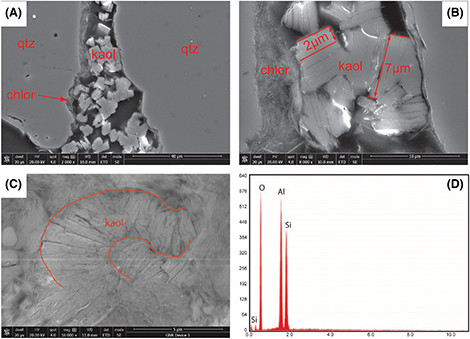
Figure 4.
Scanning electron microscopy images and energy-dispersive x-ray spectroscopy (EDS) spectrum of pore-filling kaolinite (kaol) blocks and vermicules. Chlorite (chlor) is labeled where it occurs. (A) The kaol blocks partially filling a pore. Magnification (mag) 2000×. (B) Higher mag of image (A) annotated to show crystal dimensions. The mag is 8000×. (C) The kaol vermicules. Red lines are annotations showing the curving growth habit. The mag is 16,000×. (D) The EDS spectrum taken from the area containing kaol in image (C). det = detector; ETD = Everhart-Thornley detector; GNR = graphene nanoribbon; HV = high voltage; qtz = quartz; SE = secondary electron; WD = working distance.
Figure 4. Scanning electron microscopy images and energy-dispersive x-ray spectroscopy (EDS) spectrum of pore-filling kaolinite (kaol) blocks and vermicules. Chlorite (chlor) is labeled where it occurs. (A) The kaol blocks partially filling a pore. Magnification (mag) 2000×. (B) Higher mag of image (A) annotated to show crystal dimensions. The mag is 8000×. (C) The kaol vermicules. Red lines are annotations showing the curving growth habit. The mag is 16,000×. (D) The EDS spectrum taken from the area containing kaol in image (C). det = detector; ETD = Everhart-Thornley detector; GNR = graphene nanoribbon; HV = high voltage; qtz = quartz; SE = secondary electron; WD = working distance.