The AAPG/Datapages Combined Publications Database
AAPG Bulletin
Figure
AAPG Bulletin; Year: 2021; Issue: August DOI: 10.1306/02262118214
Return to Full Text
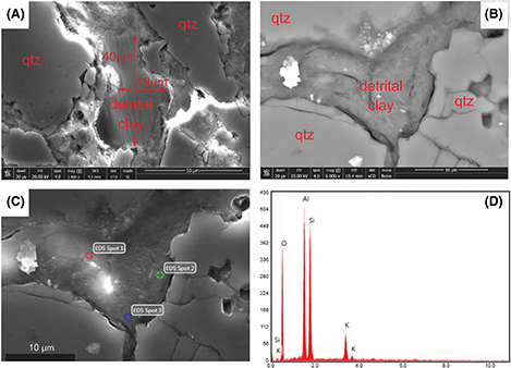
Figure 9.
Images of detrital sand-sized illitic clasts in the Cypress Sandstone. (A) Scanning electron microscopy (SEM) image of a large laminated clast. Magnification (mag) 2400×. (B) Backscattered electron image of a smaller clast between framework grains showing a fibrous texture. The mag is 6000×. (C) The SEM image collected by the EDAX image collector corresponding to (B). The clast could be interpreted as a laminated detrital clay on the basis of the areas shown in images in (B, C); however, the clast is discontinuous outside the area shown. (D) Energy-dispersive x-ray spectroscopy (EDS) spectrum typical of illitic detrital clay clasts in the Cypress Sandstone, which was taken from EDS Spot 2 on (C). The major elements of Si, Al, and K reflect the chemical composition of illite ((K, H3O)(Al, Mg, Fe)2(Si, Al)4O10[(OH)2,(H2O)]). All EDS spots taken from (C) yielded nearly identical elemental spectra. Glowing regions are the result of charging during sample–electron beam interaction. det = detector; ETD = Everhart-Thornley detector; GNR = graphene nanoribbon; HV = high voltage; qtz = quartz; SE = secondary electron; WD = working distance.
Figure 9. Images of detrital sand-sized illitic clasts in the Cypress Sandstone. (A) Scanning electron microscopy (SEM) image of a large laminated clast. Magnification (mag) 2400×. (B) Backscattered electron image of a smaller clast between framework grains showing a fibrous texture. The mag is 6000×. (C) The SEM image collected by the EDAX image collector corresponding to (B). The clast could be interpreted as a laminated detrital clay on the basis of the areas shown in images in (B, C); however, the clast is discontinuous outside the area shown. (D) Energy-dispersive x-ray spectroscopy (EDS) spectrum typical of illitic detrital clay clasts in the Cypress Sandstone, which was taken from EDS Spot 2 on (C). The major elements of Si, Al, and K reflect the chemical composition of illite ((K, H3O)(Al, Mg, Fe)2(Si, Al)4O10[(OH)2,(H2O)]). All EDS spots taken from (C) yielded nearly identical elemental spectra. Glowing regions are the result of charging during sample–electron beam interaction. det = detector; ETD = Everhart-Thornley detector; GNR = graphene nanoribbon; HV = high voltage; qtz = quartz; SE = secondary electron; WD = working distance.